Online Pet Vet Pharmacy: Best Deals Up to 40% Off + FREE Shipping on Orders $100+


Differences in heartworms in cats and dogs
Differences in heartworms in cats and dogs
The way cats get heartworm disease is very similar to how dogs get it. Maybe the only difference you can spot is that the cats are more responsive hosts to infective worms that survive to adulthood, unlike the dogs.
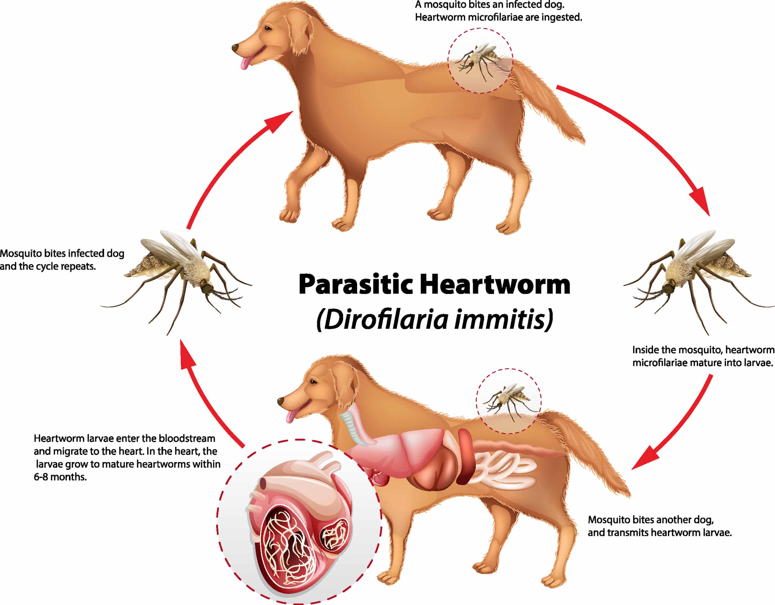
When the infective third-stage larvae (L3) penetrates the host animal, it undergoes the first molt and grows to fourth-stage larvae (L4) till the migration through host tissues is performed, transforms into an immature worm (L5) when molting a second time, and, finally, penetrates a peripheral vein. In fact, the circulatory system helps carry it and larvae flows through the heart to the caudal pulmonary arteries for further development and growth.
These immature parasites are around 2 inches in length within 100 days infection onset. When comparing cats and dogs, it’s worth mentioning that initial pulmonary artery pathology is similar. It all starts with muscular hypertrophy of main lung vessels, villous endarteritis, and a pulmonary cell infiltration to the adjacent lung parenchyma.
From now on ( a 100 day period), we are able to observe main differences in disease pathogenesis threat to feline and canine health. For the last-mentioned, the worms keep developing and progressing into adulthood until the disease is revealed. While for the cats, the majority of juvenile worms die within 3 to 4 months after infection. Due to this an acute inflammatory response and syndrome known as heartworm-associated respiratory disease (HARD) are initiated.
Such histopathological lesions can be the demonstrable evidence even in those cats that can fight off the infection and are extremely similar to those found in cats with adult worms.
The occlusive medial hypertrophy of the small pulmonary arterioles is considered to be the most commonly encountered microscopic lesion, similarly, other changes are observed in the bronchi, bronchioles, alveoli, and pulmonary arteries. Medial hypertrophy of the pulmonary arteries is often associated with Toxocara and aelurostrongylus infections. Scanned radiographic findings are similar to some forms of feline asthma or allergic bronchitis, which can result in inaccurate or missed diagnosis.
Overall prevalence rate of feline heartworm disease
An experimental infection study showed that the percentage of dogs infected with 100 L3 larvae that form 75 adult worms is extremely high, nearly 100 % of cases. Following a similar experimental model of 100 L3 larvae, but this time in cats, 1 to 10 mature heartworms will occur in 75 % of cases. Nevertheless, if conducted in a real-world scenario, the picture is quite different.
Determining the number of infective larvae in cat or dog hosts includes many features, among which we can name reservoirs and vectors of emerging infection, and temperature threshold for larvae development. Based on necropsy case studies of homeless cats that have been performed to identify adult heartworm infections, feline parasites are present at 5 to 20 % of the canine rate in the area examined.
Heartworm infection and heartworm disease exist as separate entities. The infection is when heartworms live in a host’s heart and surrounding blood vessels with a major health effect in the infected animal while the disease is a pathology unleashed as the result of current or past infections.
To sum up, everything mentioned about HARD above, immature worms are not particularly harmful, adult heartworms detected in cats do not cause life-threatening pulmonary pathological issues. To our knowledge, a necropsy study of the histopathological pattern of feline lungs was performed on a group of homeless cats that did not have adult heartworms but were heartworm antibody-positive. The study concluded that 50 % of tested cats had occlusive arteriolar hypertrophy in 20 to 40 % of arterioles.
Pilot studies in mimic abbreviated worm infections that were purposed to reproduce these lesions in the pulmonary arterioles, reported that the lesions were still observed 8months after infection.
Guidance on diagnostic testing strategy for FHWD
Most veterinarians and pet owners recognize that diagnosing heartworm disease in cats is much more tricky and complicated than in dogs. Particularly, background heartworm diagnostic testing methods used in revealing the presence of canine heartworms may not work properly for feline patients.
- Testing for Microfilariae: A common microfilariae detection test which is essentially ineffective as cats are rarely microfilameric when examined.
- Radiology: More than 50% of heartworm-positive cats have clinical radiographic signs. One of the most common signs is considered to be an enlarged on the ventrodorsal view right caudal lobar artery segment. A bronchointerstitial pattern is also one of the most common radiographic lung patterns but is not specific for feline heartworm disease. Other less frequently found signs are flattening of the diaphragm which is the sign of hyperinflated lungs, the density of parenchymal abnormalities, lobar consolidation, pleural effusion (also called water on the lungs), and, finally, pneumothorax.
- Ultrasonography:Professional ultrasound-guided check of feline adult heartworms identifies 90 % of infections; yet, the pulmonary arteries of each caudal lobe, in other words bifurcating flow, should be thoroughly followed.
- Serology: Serologic tests are now widely available and show better results than antigen and antibody tests which accuracy is often questioned for feline patients.
- Antigen tests: Infected cats typically have only one or two adult worms in their bodies; there are also infections specific for one sex only. Also, if immature worms die in a time span of 3 to 4 months after infection, it most likely results in feline heartworm disease. Due to not detecting enough early or male-only antigen in antigen tests, false-negative results may occur. On top of that, antigen-antibody immune complexes may hinder with antigen test which increases the probability of false-negative result.
Laboratory tests have demonstrated that when the sample test tube is heated up for 10 minutes to 219 ° F (104 Celsius), the complexes are induced to release antigens, that is why a higher percentage of positive test results is obtained. One of the possible diagnostic options to get better results without much doubt is now offered by most reference laboratories and is basically heat treatment testing.
Caution! The results from heat-treated samples must be used with care by healthcare professionals in order to not overinterpret them. A common example of misinterpretation is the circulation of antibodies when the actual heartworm has already died, which is not evidence of existing worms.
- There are limits to Antibody tests that show negative antibody results. This type of test can not help in tackling heartworm infections. In comparison to antigen tests which are used to identify the glycoprotein particles within the reproductive tract of adult heartworms, antibody tests show poor performance on the same antibody search.
Experimental trials of antibody tests have shown that at the first stage of the experiment, heartworm infections detection rate was 98-100 %, however, when tested on infected cats in real-world conditions, the level of accuracy was far away from high. Consequently, a high number of cats are negative to antibodies but at the same time have mature heartworms.
In one retrospective study of 50 heartworm-positive cats performed at North Carolina State University, 14 % false negatives were found, even though the vast majority of them (72 %) had signs and severe symptoms of active disease. Two other studies were successfully conducted, one shelter-based necropsy study in Texas confirmed 50 % false negative, and another study of shelter cats in Florida confirmed the rates of 11 – 68 % (again false negative) in a row of 7 different antibody tests. At least 1 of 8 tests has shown negative results for 20 out of 30 heartworm-positive cats.
Features that were compared in the studies to diagnose feline heartworm disease include repetitious antibody tests, antigen tests, and radiographs.
Client-owned cats with a primary complaint of a persistent cough were included in the first study, and shelter cats with necropsy-confirmed heartworm status and histopathology of lungs, in the second one. Both studies have highlighted that one test is not enough to provide a definitive FHWD diagnosis, plus variable capacity for diagnostic search is constantly increasing, yet, there are more FHWD than available methods to detect them.
Summary
It is essential to be aware of the number of weaknesses competent veterinarians may reveal performing antigen and antibody tests. The probability of getting an accurate diagnosis of feline heartworm disease implementing a single method is quite low unless a positive antigen test is obtained during screening or a worm shows up on an ultrasound. The animal should be tested several times in most cases, and additional tests should be performed if clinical suspicion is strong but antigen or antibody results do not detect the evidence.